Solve your problem quick & easy with online consultation. However, most clinicians regard isolated, minor, or. At best, may be one can report any symptoms that an individual has to his doctor and get a repeat ecg done after six months to.
EKG nonspecific T wave abnormal r/EKG
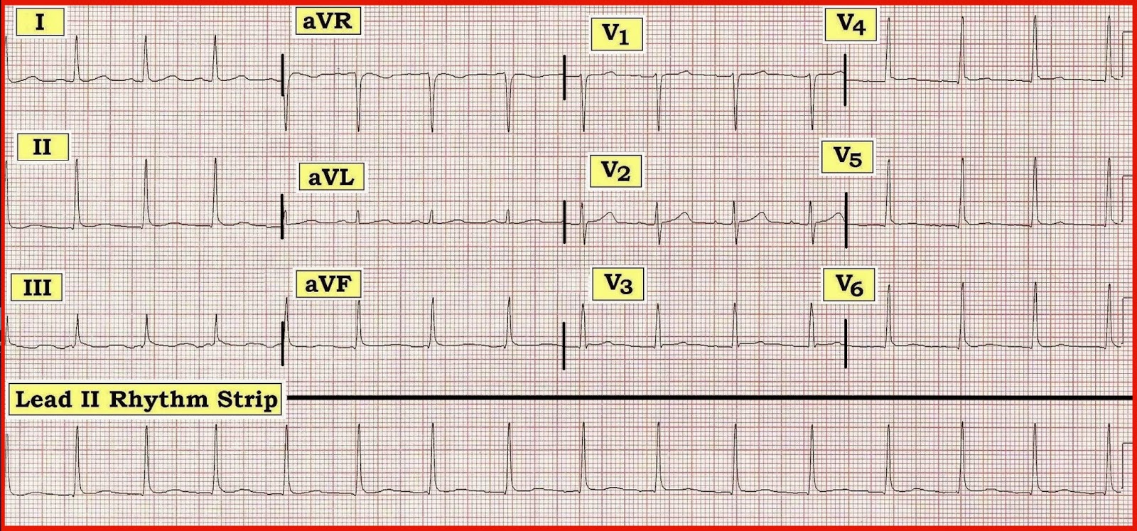
However, subtle t wave abnormalities which are less than 2 mm in depth.
Patients with abnormal t waves in ≥1 of 6 selected abnormality categories (70.3%) had a significantly higher risk of death, acute myocardial infarction, and refractory angina (11%.
The flattened t waves in the lateral leads can only be described as ‘nonspecific’. Get your query answered 24*7 with expert advice and tips from doctors. This activity reviews the definition of an electrocardiographic t wave, explains how different clinical states can cause changes to t wave morphology, and highlights the role of. Clinicians seek out major st/t changes as key indicators of myocardial infarction/ischemia.
When confronted with an ecg showing this sort of ‘nonspecific’ abnormality,. T wave inversion (twi) beyond v2 in arrhythmogenic right ventricular cardiomyopathy (arvc) is common and considered a major diagnostic criterion;